(Print) Use this randomly generated list as your call list when playing the game. There is no need to say the BINGO column name. Place some kind of mark (like an X, a checkmark, a dot, tally mark, etc) on each cell as you announce it, to keep track. You can also cut out each item, place them in a bag and pull words from the bag.
1
2
3
4
5
6
7
8
9
10
11
12
13
14
15
16
17
18
19
20
21
22
23
24
a plasma cell cancer marked by monoclonal IgG, normal anemia with rouleaux and high ESR, plus Bence Jones light chains.
Multiple Myeloma
clumped chromatin "cracked" appearance. presence of smudge cells. can make an albumin smear. many patients have no apparent symptoms.
CLL
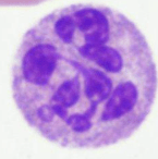
Reed sternberg cells are present. Seen in younger population, good prognosis
Hodgkin Lymphoma
Increases in this type of WBC can be seen in type 1 hypersensitivity reactions. The granules of this cell contain histamine.
Basophil
This toxic change consists of prominent large blue-black granules due to persistent staining of primary granules.
Toxic Granulation
Increases in this type of WBC can be seen in allergic and parasitic infections.
Eosinophil
<20% blasts in BM low LAP leukocytosis with left shift in WBC. predominant cells are neutrophils and myelocytes. Philadelphia chromosome.
CML
Increases in this type of WBC can be seen in tuberculosis, some autoimmune diseases, and irritable bowel syndrome. The cytoplasm has a ground-glass appearance and may contain vacuoles.
Monocyte
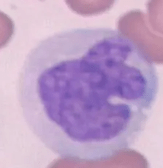
In this stage of WBC maturation, the nucleus is indented <1/2 the width of the hypothetical round nucleus. Nucleoli are absent the chromatin is coarse and clumped. Cytoplasm is pink with many secondary granules.
Metamyelocyte
Neutropenia. Lymphoblasts circulating. primarily in young children. May be CNS involvement
ALL
This is a benign, extreme or exaggerated response to an infection or stimulus accompanied by a shift to the left and toxic changes to neutrophils.
Leukemoid reaction
In this stage of WBC maturation, the production of secondary granules begins, and granulocytes can be differentiated into neutrophils, basophils, and eosinophils. The cell has an eccentric nucleus with a prominent perinuclear clearing beside the
Myelocyte
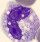
This type of B lymphocyte has abnormal cytoplasmic projections that appear “hairy” under the microscope. TRAP stain used for confirmation.
Hairy Cell Leukemia
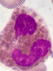
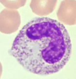
In this stage of WBC maturation, the nucleus is indented >1/2 the width of the hypothetical round nucleus. The nucleus is shaped like “C, S, or U”.
Band
This type of neutrophil has >5 lobes and is associated with megaloblastic anemia.
Hypersegmented
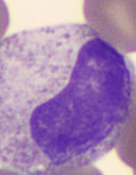
In this stage of WBC maturation, the production of primary granules begins. The chromatin is slightly condensing, and 1-3 nucleoli may be visible.
Promyelocyte
These red, staining needle-like inclusions result from the abnormal fusion of primary granules and is often seen in the cytoplasm of myeloblasts.
Auer rods
Myeloproliferative neoplasms involved with megakaryocytic cell line
ET
CD 33 and CD 13 seen in adults medium to large myeloblasts. MPO positive SBB positive
AML
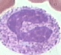
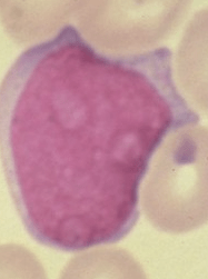
This cell is the earliest recognizable granulocyte precursor. The nucleus is round, centrally located with lightly packed chromatin and 2-5 nucleoli.
Blast
This abnormal WBC morphology is associated with chronic lymphocytic leukemia and are remnants of cells that lack any
identifiable cytoplasmic membrane or nuclear structure. Adding 22% bovine albumin to the blood sample prior to making the blood
Smudge Cell
Bone marrow fibrosis tear drops seen on blood smear. "Dry Tap"
PMF
This toxic change results in colorless areas in the
cytoplasm that indicate phagocytosis and degranulation have occurred.
Toxic Vacuolization
myeloproliferative neoplasms associated with erythroid cell line Jak2 mutation
PV